About Xenopus laevis (Daudin, 1802)
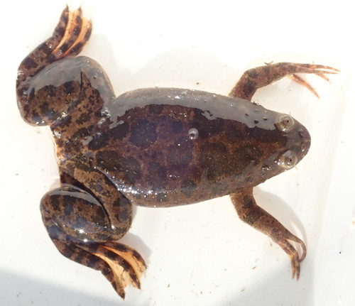
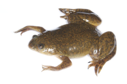
These frogs are abundant in ponds and rivers across the southeastern portion of Sub-Saharan Africa. They are fully aquatic, and typically have a mottled greenish-grey-brown coloration, sometimes with yellowish blotches, and a pale white-cream belly. African clawed frogs (Xenopus laevis) are frequently sold as pets, and are sometimes misidentified as African dwarf frogs. Albino clawed frogs are common and sold as laboratory animals. Like most amphibians, fertilization of their eggs happens outside the female's body. Among the seven known amplexus (mating position) modes for frogs, this species breeds using inguinal amplexus: the male clasps the female in front of her back legs and remains in this position until she lays eggs, then releases sperm to fertilize the egg mass. African clawed frogs are highly adaptable, and will lay eggs whenever environmental conditions are suitable. During wet rainy seasons, they travel to other ponds or puddles to search for food and new habitats. During droughts, they can burrow into mud and become dormant for up to a year. X. laevis is known to survive 15 or more years in the wild, and 25–30 years in captivity. They shed their skin every season, and consume their own shed skin. Though males lack a vocal sac, they produce mating calls of alternating long and short trills by contracting intrinsic laryngeal muscles. Females also respond vocally, making a rapping sound to accept a male or slow ticking to reject him. This species has smooth, slippery skin, with a multicolored back marked with olive gray or brown blotches. The underside is creamy white with a yellow tinge. Males and females are easily distinguished by physical traits: males are smaller and slimmer, while females are larger and more rotund. Males have black patches on their hands and arms that help them hold onto females during amplexus. Females have a more prominent cloaca, and have hip-like bulges above their rear legs where their eggs develop internally. Both sexes have a cloaca, a shared chamber that digestive, urinary, and reproductive systems empty into, which opens to the outside through the vent; in amphibians and reptiles, the vent is the single opening for all three systems. X. laevis embryos and eggs are a popular model system for a wide range of biological studies, in part because the species can lay eggs year-round when conditions allow. This animal is widely used in research due to its combination of experimental tractability and close evolutionary relationship to humans, compared to many other common model organisms. X. laevis is also notable for its role in the first widely used method of pregnancy testing. In the 1930s, South African researchers Hillel Shapiro and Harry Zwarenstein, students of Lancelot Hogben at the University of Cape Town, discovered that injecting urine from pregnant women into X. laevis induced oocyte production within 8–12 hours. This simple, reliable pregnancy test was used widely until the 1960s. In the late 1940s, Carlos Galli Mainini found in separate studies that male Xenopus and Bufo specimens could also be used to detect pregnancy. Today, commercially available hCG is injected into male and female Xenopus to induce mating behavior and breed the frogs in captivity at any time of year. Xenopus has long been an important tool for in vivo studies of molecular, cell, and developmental biology in vertebrates. The broad scope of Xenopus research also comes from the fact that cell-free extracts made from Xenopus are a leading in vitro system for studying fundamental aspects of cell and molecular biology. This makes Xenopus the only vertebrate model system that supports both high-throughput in vivo analyses of gene function and high-throughput biochemistry. Xenopus oocytes are a leading research system for studies of many biological systems, including ion transport and channel physiology. A 2001 study by Xanthos et al used oocytes to identify T-box expression earlier in vertebrate development than previously documented. While X. laevis does not have the very short generation time or genetic simplicity generally desired for genetic model organisms, it is an important model organism in developmental biology, cell biology, toxicology, and neurobiology. X. laevis takes 1 to 2 years to reach sexual maturity, and like most species in its genus, it is tetraploid. However, it has large, easily manipulated embryos, and this ease of manipulation has given amphibian embryos an important place in both historical and modern developmental biology. A related species, Xenopus tropicalis, is considered a more viable genetic model, though gene editing protocols have now been perfected for X. laevis. Roger Wolcott Sperry used X. laevis for his famous experiments describing visual system development, which led to the formulation of the chemoaffinity hypothesis. X. laevis has been used as a model organism for studies of vertebrate cardiogenesis, human congenital heart defects, and GWAS studies of congenital heart defects. Xenopus oocytes provide an important expression system for molecular biology: by injecting DNA or mRNA into the oocyte or developing embryo, scientists can study resulting protein products in a controlled system, allowing rapid functional expression of manipulated DNAs or mRNA. This is particularly useful in electrophysiology, as the ease of recording from the oocyte makes it attractive for expressing membrane channels. One challenge of working with oocytes is eliminating native proteins that could confound results, such as native membrane channels. Protein translation can be blocked or pre-mRNA splicing can be modified by injecting Morpholino antisense oligos into the oocyte (for distribution through the whole embryo) or into early embryos (for distribution only to the daughter cells of the injected cell). Extracts from X. laevis eggs are also commonly used for biochemical studies of DNA replication and repair, because these extracts fully support DNA replication and other related processes in a cell-free environment that is easy to manipulate. The first vertebrate ever cloned was an African clawed frog in 1962, an experiment that led to Sir John Gurdon being awarded the 2012 Nobel Prize in Physiology or Medicine "for the discovery that mature cells can be reprogrammed to become pluripotent". Additionally, four female African clawed frogs and stored sperm were carried on the Space Shuttle Endeavour for mission STS-47, launched 12 September 1992, to test whether reproduction and development could proceed normally in zero gravity. X. laevis also serves as an ideal model system for studying the mechanisms of apoptosis. Iodine and thyroxine stimulate widespread apoptosis of cells in the larval gills, tail, and fins during amphibian metamorphosis, and drive changes to the nervous system that transform the aquatic, vegetarian tadpole into a terrestrial, carnivorous adult frog. Stem cells from this frog were used to create xenobots.