About Symsagittifera roscoffensis (Graff, 1891)
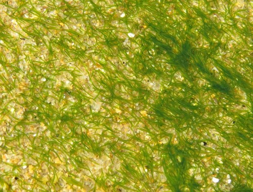
Symsagittifera roscoffensis (Graff, 1891) is an acoel flatworm that forms a photosymbiotic relationship with the microalgae Tetraselmis convolutae. Early researchers including Geddes (1879), Delage (1886) and Haberlandt (1891) observed starch and chlorophyll in the green cells inside the worm’s tissues, and suspected these cells were micro-algae, but could not formally confirm their origin or nature. In 1901, William Gamble and Frederick Keeble began studying these in hospite green cells in Roscoff, and attempted to isolate and culture them without success. In 1905, they observed non-symbiotic juveniles developed green coloration after hatching from cocoons originally laid in unfiltered seawater then transferred to filtered seawater. They hypothesized the factor that infected juveniles and produced the green color was likely located on or inside the cocoon. In a follow-up experiment, they transferred empty post-hatching cocoons originally kept in unfiltered seawater to filtered seawater for incubation. After three weeks, they observed the cocoons turned green, and green, flagellated unicellular organisms accumulated in the filtered seawater. This experiment allowed the researchers to successfully isolate these green microorganisms. Microscopic observation confirmed these cells had characteristics of micro-algae, including turning violet after iodine treatment (indicating starch presence) and showing photosynthetic activity. When non-symbiotic juveniles reared under sterile conditions were exposed to these isolated green flagellated cells, photosymbiosis was successfully induced. These foundational works confirmed that the in hospite green cells are actually flagellated microalgae that live freely in the environment, and are the infective factor that produces the green coloration absent in non-symbiotic adult worms. This work also confirmed there is no vertical transmission of symbionts from parent worms, and instead each new generation horizontally acquires symbionts from the environment. Tetraselmis convolutae belongs to the class Chlorodendrophyceae within the division Chlorophyta. In its free-living state, this alga has four flagella, a polysaccharide envelope called a theca, and a stigma (or eyespot) vacuole that holds photoreceptor molecules. T. convolutae can live freely in the water column but is mainly benthic. When it lives in hospite inside S. roscoffensis, the alga has a different phenotype than when free-living: it loses its flagella, theca, and stigma. These phenotypic changes prevented early researchers from correctly identifying the in hospite green cells as micro-algae. Although the species epithet roscoffensis means "from Roscoff", this flatworm is not endemic to Roscoff or North Brittany. Its geographical range covers the Atlantic coast of Europe, with colonies recorded from Wales to southern Portugal. Adult S. roscoffensis reach 4 to 5 mm in length. The anterior head region holds a gravito-sensing statocyst (or otolith) that allows the worm to orient in space, and produces negative geotropism: a mechanical stimulus against the wall of a tube holding the worms triggers active diving to the bottom of the tube. Two photoreceptors flank the statocyst; light perception lets the worm move toward illuminated environments, a trait called positive phototropism. This adaptation likely increases the chance of encounters between free-living microalgae and non-symbiotic juveniles, which also have positive phototropism. When exposed to varying light intensities, S. roscoffensis tends to move and expose itself to higher light intensities than those where free-living microalgae achieve optimal photosynthesis. Experimental work also shows that when given a choice, photosymbiotic worms avoid extreme light conditions that are either too weak or too strong. In their natural environment, temporary burying is thought to let worms escape intensities that are too high and would cause photoinhibition. This species has a central brain and peripheral nervous system. If the anterior head region containing the brain is amputated, the worm can regenerate the entire central nervous system in around twenty days, and normal behavior recovers alongside regeneration. Different biological functions regenerate at different speeds: phototropism, linked to photoreceptor regeneration, recovers quickly, while geotropism, linked to statocyst regeneration, takes several weeks to recover. S. roscoffensis has no circulatory blood system, so oxygen diffuses passively through its tissues. Some of this oxygen is produced by the photosynthetic activity of in hospite algae. The worm’s body surface is covered in abundant cilia and dotted with numerous mucus-secreting glands. Mucus forms a physical network that allows the worms to move through seawater seeps. In situ study of S. roscoffensis behavior confirms that the worm can only move horizontally by building a naked-eye invisible support structure made from a matrix of its own secreted mucus. The worms do not slide directly on sand, and instead give the impression of sliding on an invisible surface. The study’s author hypothesized that moving above the sandy substrate lets the worms receive more light via reflected light rays, allowing more photosynthesis from symbiotic algae and greater nutrient transfer to the worm. Secreted mucus also acts as a biofilm interface between the worm and its environment. Specific bacterial populations grow in and are hosted by the mucus, and are thus closely involved in the worm’s biology. This three-way association of animal, micro-algae, and bacterial consortium is a well-studied example of the holobiont paradigm, which holds that an organism is a complex, dynamic association with microbial communities that are necessary for the organism’s development, growth, and to some degree its survival. S. roscoffensis never has a digestive system at any stage of its development. It has a ventral opening considered a mouth that lets it ingest, but not digest, the microalga T. convolutae. Ingested microalgae enter a digestive syncytium, where future in hospite microalgae first become vacuolated, losing their flagella and theca, then transit to finally locate under the worm’s epidermis. The algae are not internalized into worm cells, instead residing between and in contact with worm cells. S. roscoffensis has a muscular system made of a complex network of transversal, longitudinal, circular, and dorsal-ventral muscle fibers. This worm is a hermaphrodite that cannot self-fertilize, so it must mate with a partner to reproduce. Mature sperm are produced at the posterior end of the animal. After mating, partner sperm is stored in a spermatheca, which connects to oocytes via a single canal. Oocytes are fertilized by stored partner sperm. Each gravid individual uses its abundant secreted mucus to produce a transparent cocoon, where fertilized oocytes are released. Embryo counts vary, and can reach up to twenty, and all embryos develop synchronously. Under laboratory rearing conditions, juveniles hatch after 4 to 5 days, escape the cocoon, and begin searching for their photosynthetic symbiont. If juveniles do not ingest the microalgae, non-symbiotic laboratory juveniles die after around 20 days.