About Calvatia craniiformis (Schwein.) Fr. ex De Toni
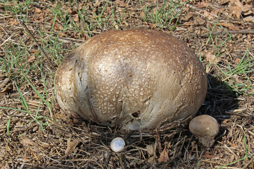
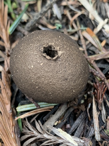
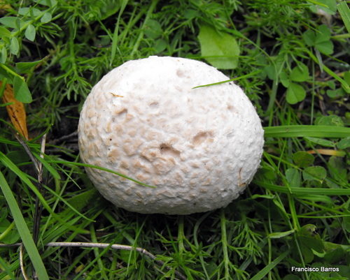
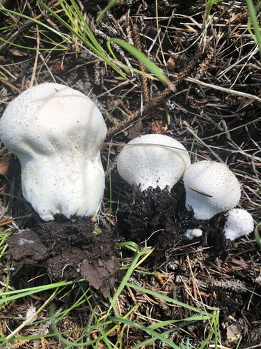
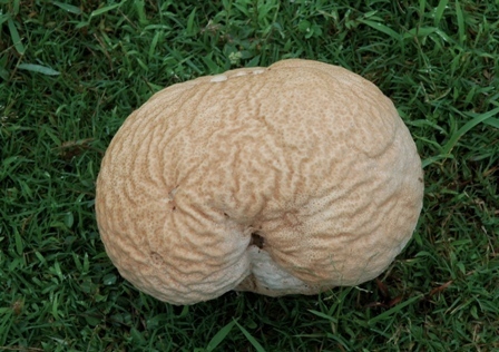
Fruit bodies of Calvatia craniiformis measure 6–20 cm (2.4–7.9 in) tall by 8–20 cm (3.1–7.9 in) wide. Their shape ranges from pear-shaped, flattened-spherical, obovate (roughly egg-shaped), to tunicate (inverted cone-shaped). At the base, a thick, often crumpled structure attaches to a cord-like, well-developed rhizomorph that is frequently encrusted with surrounding soil. When cut longitudinally, the rhizomorph shows three distinct tissues: an outer cortex, a subcortical layer, and a central core. The thin, fragile outer layer of the peridium (exoperidium) is whitish-gray to gray, starting smooth before developing an areolate texture split by cracks into separate patches. The base extends one-third to halfway up into the puffball as a tapering, pointed columnella. The internal spore-bearing tissue (gleba) is initially white, turns yellow-green, and becomes brownish-green in older specimens with mature spores. Spores of this species are spherical, translucent (hyaline), 2.5–3.4 μm in diameter, with thick walls, a short tubelike extension called a pedicel, and ornamented with small, roughly evenly spaced wartlike projections called verrucae. Capillitial threads are long, hyaline, branched, 2.4–4 μm thick, septate, and occasionally bear pits on their walls. The exoperidium is made of thick-walled inflated hyphae mixed with spherical sphaerocysts, while the inner endoperidium consists of tightly interwoven thick-walled hyphae. In the rhizomorph, hyphae in the central core are several times thicker than hyphae in the surrounding subcortex. Light microscopy generally cannot distinguish the spores of C. craniiformis from those of C. rubroflava and C. gigantea, but electron microscopy reveals distinct ornamentation for each species: C. craniiformis has small, well-separated verrucae up to 0.2 μm tall with rounded tips, while C. gigantea has larger verrucae up to 0.4 μm tall with more irregular arrangement. Calvatia craniiformis is generally classified as a saprobic species, but a 1966 publication reported that it forms ectomycorrhizae with American sweetgum (Liquidambar styraciflua) in controlled laboratory conditions. A Chinese study found that C. craniiformis readily forms mycorrhiza with poplar seedlings in unsterilized soil, but not in sterilized soil. Later research failed to establish a similar mycorrhizal association between C. craniiformis and Pinus ponderosa. Commonly called brain puffball, this fungus grows singly or in groups in fields, open woods, hardwood forests, and wet areas. Its distribution includes Asia (recorded in China, India, Indonesia, Japan, Malaysia, and South Korea), Australia, and North America (eastern and southern United States, and Mexico). In Michigan, it is one of the few macrofungi that occurs regularly in black locust plantations. Its fruit bodies act as a food source for several fly species. Calvatia craniiformis is an edible species. Young puffballs with firm, white gleba have a mild odor and pleasant taste. Early 20th-century mycologist Charles McIlvaine noted over a century ago that even the slightest shift to yellow in color makes the puffball bitter. It is versatile for cooking, as it absorbs flavors well. In the United States, the Ojibwe people used the powdery mature gleba as a hemostatic agent to stop nosebleeds, by inhaling the spore powder through the nostrils. We now know this practice can cause lycoperdonosis, a pulmonary disease with symptoms similar to pneumonia. This puffball is also used as a hemostatic agent in Chinese and Japanese folk medicine.