About Amanita ravenelii (Berkeley & Broome) Saccardo, 1887
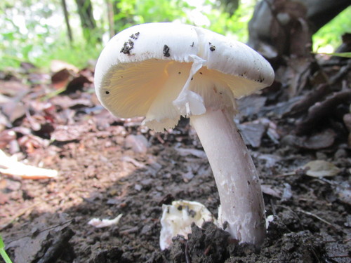
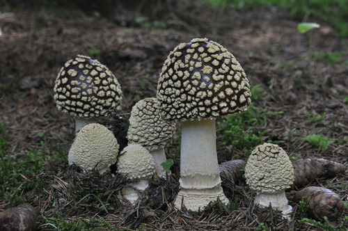
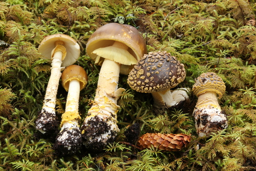
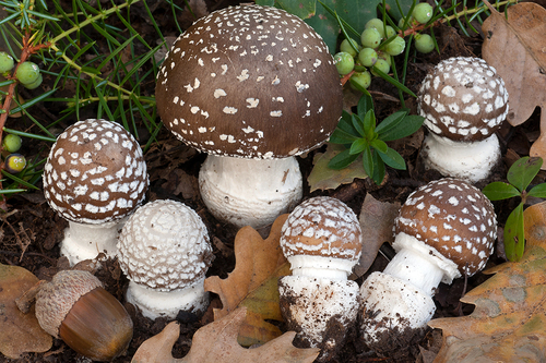
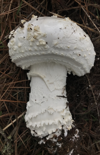
The scientific name of this fungus is Amanita ravenelii (Berkeley & Broome) Saccardo, 1887. Its cap measures 8–17 cm (3–6+1⁄2 in) wide. It starts as hemispherical to nearly round, and matures to become convex to flattened. The cap is fleshy, colored white to yellowish-white, and usually dry, occasionally becoming slightly sticky with age. Remnants of the universal veil remain as a pale yellow to brownish-orange layer that breaks into crowded, coarse warts that are conical to truncate-conical. These warts grow up to 6 mm (1⁄4 in) wide and 4 mm (1⁄8 in) high, and become more scale-like near the cap margin as they age. The cap margin is non-striate, with no grooves, and appendiculate, with partial veil remnants hanging along its edge. The gills do not attach to the stem, are crowded, moderately broad, and colored yellowish-white to pale yellow. Short gills called lamellulae do not reach all the way to the stem, and are somewhere between abruptly truncated and gradually tapered. The stem measures 10–25 cm (4–10 in) long and 1–3 cm (1⁄2–1 in) wide, and tapers slightly near its apex. It is solid, not hollow, colored white to pale yellow, and covered in soft woolly tufts of hair or fibrils. It has a large middle-swollen basal bulb that extends up to 5.5 cm (2 in) into the ground. The partial veil is yellowish-white to pale yellow, and forms a thick, woolly, delicate ring that quickly falls off. Universal veil remnants remain at the stem base as thick downward-curved scales that often form irregular rings. The flesh is firm, and colored white to pale yellow. Mushroom tissue has an odor described as chlorinated lime (bleaching powder), chlorine, "old tennis shoes", or "old ham". Microscopically, the spores are ellipsoid, occasionally ovoid or obovoid, thin-walled, hyaline, and amyloid, and measure 8–11 by 5.5–7.5 μm. Spore deposits are white. The spore-bearing basidia measure 40–65 by 7–11.5 μm, are four-spored, and have clamps at their bases. Cheilocystidia, cystidia on the gill edge, are occasionally found as small club-shaped cells 15–35 by 10–15 μm, growing on thin-walled hyphae that are 3–7 μm in diameter. The cap cuticle is not clearly distinct from cap tissue, and is made of thin-walled, interwoven hyphae 2.5–9 μm in diameter. The universal veil tissue on the cap is made up of roughly parallel, erect rows of roughly spherical, and ellipsoid to broadly ellipsoid cells up to 78 by 65 μm, plus spindle- to club-shaped cells up to 125 by 30 μm. These spindle- to club-shaped cells are terminal or arranged in short terminal chains, and grow from moderately abundant, thin-walled, branched, interwoven, sometimes nearly coralloid hyphae 3–9.5 μm in diameter, with a small number of scattered 5–12.5 μm diameter oil-containing oleiferous hyphae. Hypha distribution at the stem base matches that on the cap, but has more filamentous hyphae. Clamp connections are present. Amanita ravenelii is widely distributed across the southeastern United States, where it occurs occasionally to frequently from late summer to autumn, between August and November. It has been collected from the U.S. states of Maryland, North Carolina, South Carolina, Indiana, Tennessee, Virginia, and Kentucky. It has also been reported growing in northern Baja California, Mexico, and in the Argentine provinces of Tucumán, Buenos Aires, and Rio Negro. It is a mycorrhizal fungus, meaning it forms mutualistic associations with shrubs and trees. Mushrooms grow on the ground singly, scattered, or in groups in mixed coniferous and deciduous forests. While the specific tree associations preferred by A. ravenelii are unknown, Amanita species from section Lepidella generally associate with diploxylon pine (pines in subgenus Pinus), oak, and hickory. The edibility of species in Amanita subgenus Lepidella is variously described as unknown, not recommended for eating, or poisonous.